Interpreting pacemaker ECGs
Assessing pacemaker function requires knowledge of the mode of pacing, and careful analysis of ECG tracings. Most modern devices are capable of transmitting ECG tracings continuously to cloud-based platforms, which enables the clinician to examine intracardiac ECGs at any time. However, most clinicians who encounter patients with pacemakers only have access to conventional surface ECGs. Being able to assess pacemaker function and perform troubleshooting should be considered a basic clinical skill.
Pacing activity may be visible or invisible, depending on e.g the type of pacemaker, intrinsic cardiac activity, etc. The cardinal manifestation of pacing on surface ECG is the stimulation artifact (Figure 1). In atrial pacing, the stimulation artifact precedes the P-wave. In ventricular pacing, the stimulation artifact precedes the QRS complex. Two artifacts are seen if both chambers are paced. The stimulation artifact is larger in unipolar pacing, as compared with bipolar pacing. The latter yields a discrete stimulation artifact, which may be visible in one or a few leads.
In addition to stimulation artifacts, ventricular pacing yields wide QRS complexes with LBBB morphology (i.e left bundle branch block appearance). This is explained by the fact that, as in LBBB, the left ventricle receives the depolarizing impulse from the right ventricle (where the pacemaker delivers the pulses). The depolarizing wave spreads outside the conduction system, which is considerably slower, as compared with impulse transmission within the conduction system (His-Purkinje network).
Assessing pacemaker function
Base rate
The base rate is the lowest heart rate allowed by the pacemaker; intrinsic cardiac activity below the base rate will trigger pacing. The base rate is usually set to 60 beats/min. The base rate is virtually always >50 beats/min, meaning that any heart rate below 50 beats/min is most likely not paced. An intrinsic heart rate faster than the base rate should inhibit the pacemaker.
P-waves
The appearance of the P-wave depends on where the atrial lead is fixed. Typically, the atrial lead is fixed next to the right atrial appendage, or atrial ceiling, which yields P-waves similar to those seen during normal sinus rhythm (i.e, positive P-wave in lead II). If the atrial lead is placed distally in the atrium, activation may proceed in the opposite direction, which results in negative (retrograde) P-waves in lead II.
QRS Complex
QRS morphology also depends on where the pacing stimulus is delivered. Typically, the lead tip is fixed apically in the right ventricle; activation starts in the right ventricle and spreads slowly to the left ventricle. As mentioned above, this is similar to the situation in left bundle branch block (LBBB), which explains why paced QRS complexes are similar to the QRS morphology during LBBB.
Stimulation in other regions of the ventricle may result in a different QRS morphology. If the lead tip is fixed in the septum, the impulse may actually enter the conduction system (His-Purkinje network), which results in rapid impulse transmission and thus shorter QRS duration (as compared with apical pacing).
Because ventricular pacing results in abnormal depolarization, repolarization will also be abnormal, resulting in discordant ST-T segments (i.e the QRS complex and T-wave display opposite directions).
Below follows ECG tracings demonstrating these aspects.
Figure 2. Atrial pacing with normal conduction to the ventricles via the AV system. The ventricles are depolarized via the His-Purkinje network, resulting in normal QRS duration.
Figure 3. Spontaneous atrial activity is sensed by the atrial lead and triggers ventricular stimulations. The QRS complex is wide due to ventricular depolarization proceeding outside the conduction system.
ECG in biventricular pacing (CRT)
In biventricular pacing, stimulation occurs in both the right and left ventricle. With simultaneous atrial spacing, a total of three stimulation artifacts can be seen on surface ECG. Stimulation of the right and left ventricle need not occur exactly at the same time. The purpose of biventricular pacing is to synchronize ventricular contraction. This mode of pacing, referred to as cardiac resynchronization therapy (CRT), reduces morbidity and mortality in chronic systolic heart failure with a wide QRS complex. CRT does not, however, reduce morbidity and mortality in patients with QRS duration of less than 130 msec (1-4).
Fusion and pseudofusion
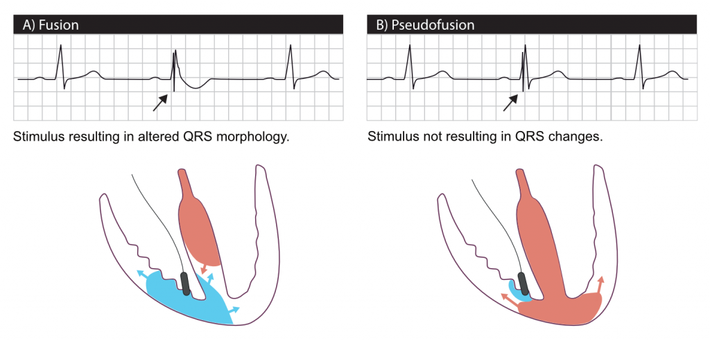
Fusion implies that the ventricle is simultaneously depolarized by the pacemaker stimulus and the intrinsic impulse passing through the His-Purkinje system. Fusion occurs if the pacemaker fails to sense intrinsic ventricular depolarization. It can also occur if the pacemaker senses the intrinsic depolarization too late.
Because the ventricle is activated both by the pacemaker stimulus and the intrinsic impulse, the QRS morphology resembles a fusion between a normal beat and a paced beat (Figure 8A).
Pseudofusion occurs in the same situations as fusion, but the depolarization from the pacemaker stimulus fails to spread through the myocardium (because it is refractory after conducting the intrinsic impulse). A stimulation artifact is seen, but the QRS complex is not affected (Figure 8B).
Acute myocardial infarction in a patients with pacemakers
Atrial pacing does not affect the QRS and ST-T segment. Thus, atrial pacing does not affect the interpretation of myocardial ischemia on ECG.
Ventricular pacing, however, results in a wide QRS complex and secondary ST-T-changes, which complicates detection of ischemia. As in left bundle branch block, these secondary ST-T changes may mask or mimic acute myocardial ischemia. There are three methods to approach this problem:
- Temporarily inactivate the pacemaker, if the patient has intrinsic cardiac activity. This potentially allows for examination of the ST-T segments during normal depolarization and repolarization. Note that switching off the pacemaker is a risky procedure, and cardiac memory may result in persisting ST-T changes even during normal ventricular depolarization. Cardiac T-wave memory implies that ST-T changes seen during pacing persist for a period after pacing is inactivated.
- Compare the current ECG tracing with previous tracings, in order to evaluate ST-T changes. Such changes may suggest ongoing ischemia.
- Use the Sgarbossa criteria, although they have not yet been validated for paced rhythms.
Pacemaker malfunction, including ECG interpretation, is discussed in the next chapter.
References
- Ruschitzka et al (N Engl J Med 2013; 369:1395-1405) – Cardiac-Resynchronization Therapy in Heart Failure with a Narrow QRS Complex
- Goldenberg et al (N Engl J Med 2014; 370:1694-1701) – Survival with Cardiac-Resynchronization Therapy in Mild Heart Failure
- Tang et al (N Engl J Med 2010; 363:2385-2395 – Cardiac-Resynchronization Therapy for Mild-to-Moderate Heart Failure
- Moss et al (N Engl J Med 2009; 361:1329-1338) – Cardiac-Resynchronization Therapy for the Prevention of Heart-Failure Events.


